Links are provided to the websites and viewers are encouraged to visit the original sites which have more pictures, discussions, references to papers, job opportunities, software, lists of staff, etc.
Photodynamic therapy (PDT) is a light-activated chemotherapy which uses light to activate a photosensitizing agent that causes oxidative damage to cells. PDT is being used for cancer therapy and is being developed for other therapies such as clearing psoriasis, alleviating arthritis, and destroying virus.
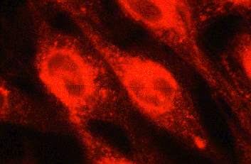 CLICK HERE to expand figure. | Accumulation of fluorescent photosensitizer in cancer cells.Photosensitizing agents are often fluorescent which allows visualization of how the agent distributes in cells. In this case, Photofrin (trademark, Quadralogic Technologies Inc.) appears as red fluorescence that has accumulated in membranes and mitochondria of cells. |
| Univ. of Texas M. D. Anderson Cancer Center Prepared by Xian-Yan He and Steven Jacques. |
 CLICK HERE to expand figure
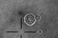 CLICK HERE to expand figure
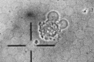 CLICK HERE to expand figure
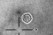
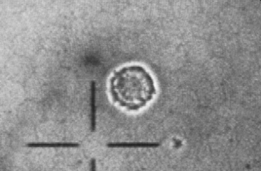
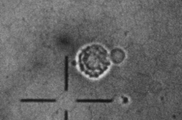
MTF7 cancer cells were incubated in ALA (delta-aminolevulenic acid, a precursor for porphyrin synthesis) for 20 hrs which caused the cells to accumulate PPIX (protoporphrin IX, the final step in porphyrin synthesis before adding heme in the synthesis of hemoglobin). PPIX is a potent photosensitizer. An argon laser (blue light, 488 nm) was directed down through the microscope to cause activation of the PPIX. These pictures were taken with a cutoff filter blocking the argon laser light and video taped on a recorder. The formation of blebs occured within the first two minutes of irradiation and is attributed to oxidative damage to cell membranes. Figure 1: before activating PPIX. Figure 2: after about 1 minute of blue light activation. Figure 3: after about 2 minutes of blue light activation. | Killing a cancer cell with PDTThis sequence of pictures shows a cancer cell being killed with PDT within the first 2 minutes of light activation of the photosensitizer PPIX which has accumulated in the cell after incubation in media with precursor ALA. The PDT causes oxidative damage to cell membranes which yields the membrane "blebs" in Figures 2 and 3. |
| Univ. of Texas M. D. Anderson Cancer Center Prepared by Kunio Awazu and Steven Jacques. |
NewsEtc. Index
OMLC home page