A reflectance-mode confocal microscope was built which delivered argon ion laser light (488 nm wavelength, incident power Po < 10w mW) and collected the backscattered 488 nm light. (The fluorescence excited by the argon ion laser was also collected in a separate channel, but that is another story.) The following Monte Carlo simulations illustrate the experiment.
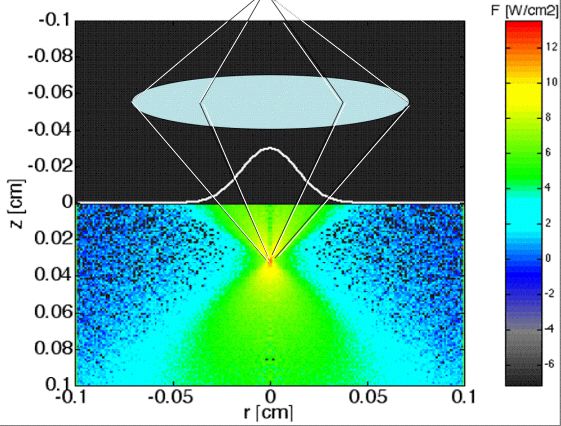
Confocal microscope collects light through a pin hole that only accepts photons scattered from the focus. (Above is only a schematic illustration. True dimensions were lens N.A. = 0.90, working distance = 3mm, focus movement within upper 100 μm of tissue used for analysis.)
Experiment scans the focus of the microscope down into the tissue to yield signal as a function of the depth of the focus, zf. (Moving black lines indicate movement of the focus.) The figure shows the light transport, T(r,z) [W/cm2 per W incident] or [cm-2], into the tissue. Colorbar shows log10(T).
When the focus is within one transport mean free path, mfp' = 1/(μs(1-g)), of the surface, the T at the focus falls exponentially versus zf. However, when attempting to focus to deeper depths, a simple diffuse light pattern develops and dominates the signal. Therefore, this report analyzes confocal signals generated by focusing within one mfp' of tissue.