The objective is derived from the need for a reliable absorption spectrum of lipids for component analysis of in vivo tissue spectra. NIR in vivo spectroscopy enables to derive the concentration of the key tissue constituents absorbing in the 600-1100 nm range, that is oxy- and deoxyhemoglobin, water and lipids. Yet, although the first three constituents are already well characterized in literature, quite few data are available on mammalian lipids. In the present proceeding we report the absorption spectrum of a clear purified oil obtained from pig lard. Absorption coefficients were measured with time resolved and spatially resolved diffuse reflectance spectroscopy techniques. At temperatures of 37°C and higher it is a clear transparent liquid thus suitable for collimated transmission measurements. In total three independent measurement techniques were employed the determine the absorption coefficients of mammalian lipids.
Materials & Methods
5 kg of pig lard was divided in pieces of 1 cm3 and placed in water with a temperature of 90°C. After a while a thin layer of pure oil from the lard formed on top of the water, and was removed by a spoon and placed in a separate container. This process was continued for 6 hours until the oil secretion process had slowed down. The substance obtained still contained water and other visible tissue structures. After cooling the oil had solidified into a pure white grease that could be separated easily from the remaining water and a gelatinous substance. The solid oil was then heated once more to 80°C and filtered twice in liquid state. After that the oil was heated again to 80°C and poured onto a filter containing Sodium sulphate to remove the last traces of water. Finally the oil was placed in a centrifuge for 30 minutes at 1000 rpm at a constant temperature of 70°C. The bottom of the reaction tubes still contained some remaining sediment and the pure oil was separated by pipette from the tube, resulting into 250 ml of oil that is visually clear at temperatures of 37°C and up.
Light from a 100 W quartz tungsten halogen lamp is coupled into an optical fiber leading to a cuvette holder and collimated to a beam of approximately 2mm diameter. Three different cuvettes were used (10, 20 and 50 mm). The collection of the transmitted light was performed by an integrating sphere with a collection port much larger that the beam diameter to compensate for differences in beam diameter caused by the divergence of the light beam and the different cuvette path lengths. The collected transmittance was spectrally projected onto a 16-bit 256-1024 pixel CCD camera cooled to -30°C Cuvette holder plus integrating sphere were placed in an oven. Measurements were performed at constant temperatures of 37, 60 and 80°C. For each cuvette path length three sequential transmission and background measurements were performed.
Results
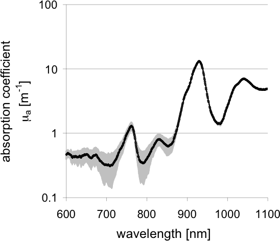
The graph below shows the absorption coefficient corrected for scattering contribution versus wavelength. The error bars represent the standard deviation over the 2 temperatures i.e. 37, 60°C
Data is available for the absorption coefficient of fat versus wavelength. Note that this has been slightly processed (by Scott Prahl) so that wavelengths are specified at whole numbers and so that absorption values have only three digits of accuracy. The original data is also available. Further details are available in the conference proceedings.